Echocardiogram / Stress Echo

An Echocardiogram is a sonogram to look at the heart. It uses sound waves just like the sonograms for pregnancy, only it uses frequencies of sound that work better in the chest. It is just like the Color Doppler Radar that you see on the weather only at frequencies of sound that work better in the chest.
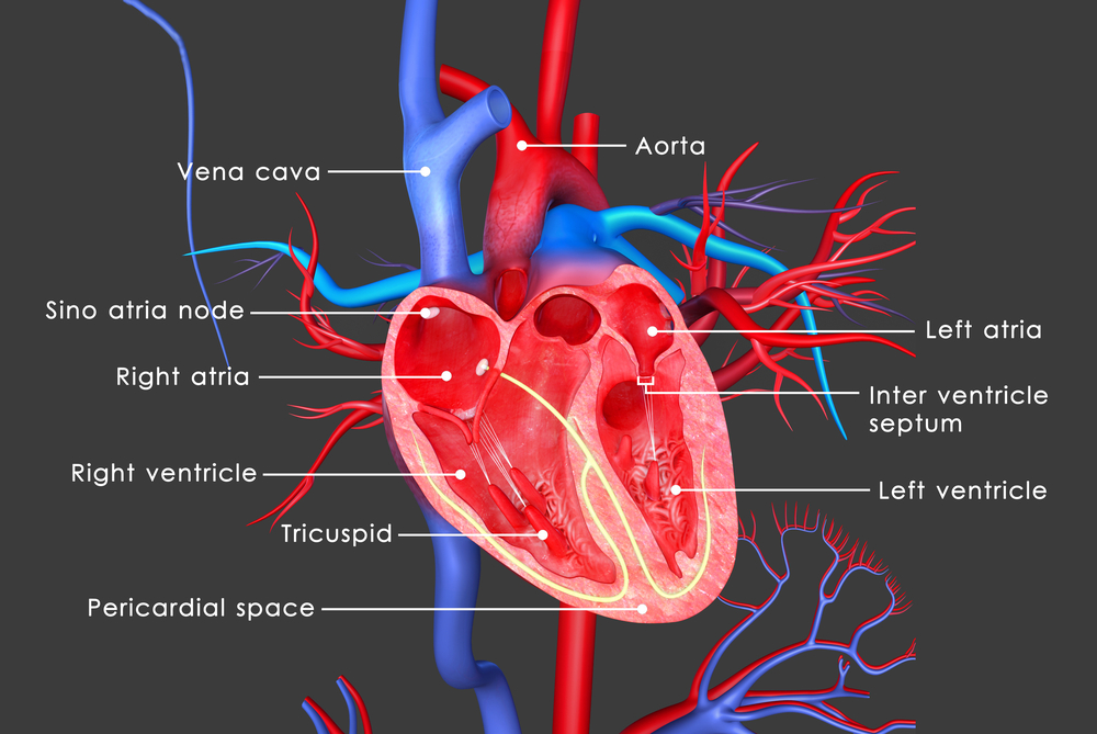
By looking at the heart you can see dimensions of the various parts, valves, chambers, and muscle. You can also see how they function and watch the blood flow through the heart, with red being one direction blue being the opposite direction. This is a very visual test and how the heart functions can be seen right on the screen. You will be asked to lie on your left side and, as with any ultrasound, a little gel and an ultrasound transducer will be placed on your chest. You just need to lie still and watch (if you want to). The technician will measure the valves; chambers, muscle and blood flow through the heart from as many different sites as possible. The technician will direct you to move or roll as necessary and the last view will be flat on your back looking directly under the ribcage to view the heart.
This will be recorded for your physician to see and then give you the results. These results can then be measured again in the future to see changes and effectiveness of therapies; you just compare the pictures and measurements. No prior preparation is needed. It is painless, remarkably easy, highly reliable, and very accurate.
A Stress Echocardiogram is an echocardiogram followed by a stress test followed by another echocardiogram as fast as possible afterwards. A stress test is being hooked to an EKG (the sticky patches placed on your chest and the wires connected to the patches that make the waveform you see and blips your hear on the EKG monitor) and walking on a treadmill to increase the workload on the heart or if walking on a treadmill is not an option, being hooked to an EKG and having a drug administered to increase your heart rate and heart workload. You are combining the stress test and the echocardiogram so you can actually watch and compare how the heart performs normally and under increased workloads.
After your stress test the technician will have you as fast as possible lie down on your left side and will direct you when to move or may direct you to stop breathing for 15 seconds so the lungs don’t obscure the picture. The before stress test images and after stress test images are then compared to see how your heart functions under more demanding circumstances.
Your physician is there for this exam and watches while it is performed and recorded for your medical record. Your physician can then give you the results immediately and let you know how your heart is performing. The results of both tests can then be measured again in the future to see changes and effectiveness of therapies; you just compare the pictures and measurements. No prior preparation is needed. It is painless, remarkably easy, highly reliable and very accurate.